If you love lifting heavy weights, you have probably encountered the frustrating reality of knee discomfort. Whether it manifests as an annoying ache right below the kneecap during heavy leg presses, a sharp twinge at the bottom of a deep squat, or persistent stiffness when walking downstairs the morning after leg day, knee problems can quickly derail your strength and hypertrophy goals. Many lifters respond by abandoning squats entirely, wrapping their joints in tight neoprene sleeves, or resigning themselves to the myth that lifting heavy weights inevitably destroys your cartilage.
The human knee is not a fragile hinge joint doomed to wear out under load; it is a highly adaptable biological structure capable of remodeling, strengthening, and thriving under progressive mechanical tension. Achieving lasting knee durability does not require avoiding knee-dominant exercises. Instead, it demands an understanding of tendon physiology, joint contact stresses, and the neuromuscular stabilizers that protect the joint capsule. By applying targeted isometric tendon loading, restoring full joint articulation, and strengthening the surrounding muscular architecture, you can permanently eliminate knee irritation, restore deep squat depth, and build joints that withstand decades of hard training.
The Anatomy and Biomechanics of the Knee Joint
To build resilient, indestructible knees, you must first understand the complex mechanical junction that forms the knee joint. The knee is not a simple hinge that moves solely in one plane; it is a complex modified condylar synovial joint comprised of two primary functional articulations:
- The Tibiofemoral Joint: The weight-bearing articulation between the rounded condyles of the distal femur and the flat tibial plateau of the shin. This joint carries the vast majority of axial compressive load during standing, running, and lifting.
- The Patellofemoral Joint: The articulation between the posterior articular surface of the patella (kneecap) and the trochlear groove of the anterior femur. The patella functions as a mechanical pulley, increasing the moment arm and mechanical leverage of the quadriceps muscle group by roughly thirty to fifty percent.
Surrounding and stabilizing these bony articulations is an intricate web of passive and active restraints. The primary passive stabilizers include the anterior cruciate ligament (ACL) and posterior cruciate ligament (PCL), which prevent excessive anterior and posterior translation of the tibia relative to the femur. The medial collateral ligament (MCL) and lateral collateral ligament (LCL) resist valgus (inward collapse) and varus (outward bowing) torques, while the medial and lateral menisci—two crescent-shaped fibrocartilaginous pads—absorb shock, distribute compressive stress across the tibial plateau, and enhance joint congruency.
The active muscular stabilizers dictate how forces travel through the joint during movement. The four heads of the quadriceps femoris—the rectus femoris, vastus lateralis, vastus intermedius, and vastus medialis (specifically the oblique fibers, or VMO)—converge into the thick quadriceps tendon, which encases the patella and continues downward as the patellar tendon (patellar ligament), anchoring directly into the tibial tuberosity. On the posterior side, the biceps femoris, semitendinosus, and semimembranosus (the hamstrings), along with the gastrocnemius (calves) and popliteus, provide posterior joint stability and protect the cruciate ligaments during deep knee flexion.
Tendon Physiology: Why Connective Tissue Breaks Down
The vast majority of non-traumatic knee pain experienced by strength athletes does not originate from torn ligaments or worn-out bone; it stems from patellar tendinopathy (often colloquially termed "jumper's knee") or patellofemoral pain syndrome (PFPS). To understand why these conditions develop, you must look at the biological differences between muscle and dense fibrous connective tissue.
Skeletal muscle tissue is an extraordinarily vascular organ. A vast capillary bed constantly bathes muscle fibers in oxygen, systemic hormones, glucose, and circulating amino acids. When you train a muscle group to failure, micro-tears in the myofibrils initiate an immediate inflammatory response followed by a surge in muscle protein synthesis, repairing the muscle belly within forty-eight to seventy-two hours.
Tendons and articular cartilage, by contrast, are notoriously hypovascular structures. The central portion of the patellar tendon receives only a tiny fraction of the capillary blood supply seen in adjacent muscle bellies. Instead of relying on systemic perfusion, chondrocytes (cartilage cells) and tenocytes (tendon cells) rely primarily on synovial fluid diffusion and mechanical fluid pumps driven by cyclic compression and decompression during movement.
Because tenocyte cellular turnover and collagen synthesis occur at a substantially slower rate than myofibrillar protein synthesis, muscular strength frequently outpaces connective tissue capacity. When a lifter rapidly increases squat frequency, adds heavy loading too quickly, or jumps into aggressive plyometrics, the quadriceps muscles have no trouble generating the necessary force. However, the patellar tendon experiences accumulated micro-strain that outpaces its collagen remodeling rate.
When overloaded continuously without adequate recovery, the tendon enters what sports scientists describe as the tendinopathy continuum:
- Reactive Tendinopathy: An acute, non-inflammatory proliferative response to acute overload, where the tendon ground substance swells with bound water molecules to protect its structural integrity.
- Tendon Disrepair: The extracellular matrix begins to break down, collagen fibers lose their parallel alignment, and abnormal capillary and nerve ingrowth (neovascularization) occurs, producing chronic localized sensitivity.
- Degenerative Tendinopathy: Areas of localized tenocyte cell death and disorganized matrix, leaving the tendon structurally compromised and prone to persistent pain under load.
True joint durability protocols intervene early on this continuum, utilizing targeted mechanical loading to stimulate mechanotransduction—the cellular process by which physical forces are converted into biochemical signals that drive type-I collagen synthesis and restore tendon matrix stiffness.
The Isometric Breakthrough: Rapid Tendon Pain Relief
For decades, the standard medical advice for knee pain was complete rest, anti-inflammatory medications (NSAIDs), and ice. Modern sports science has thoroughly debunked this passive approach. Complete rest does not heal a painful tendon; it actually causes the tendon to decondition, reducing its load capacity and rendering it even more vulnerable when you resume training.
The true breakthrough in knee tendon durability came from landmark research led by sports medicine researcher Dr. Ebonie Rio and colleagues.[1] The researchers investigated how different exercise modalities affected patellar tendon pain and central nervous system inhibition in athletes with patellar tendinopathy.
When a tendon is in pain, the central nervous system places a protective "governor" over the quadriceps muscle, known as arthrogenic muscle inhibition. The brain actively reduces high-threshold motor unit recruitment to prevent the lifter from producing high forces through the painful joint. Dr. Rio's research demonstrated that performing heavy, sustained isometric contractions (five sets of forty-five-second holds at approximately seventy percent of maximal voluntary contraction) produced an immediate, profound analgesic effect, dramatically reducing tendon pain for up to forty-five minutes while simultaneously releasing cortical inhibition in the motor cortex.[1]
Unlike dynamic concentric or eccentric movements—which involve rapid stretch-shortening cycles, high peak forces, and compressive tendon friction against bone—isometric muscle actions apply steady, static tension. This continuous tension triggers a phenomenon known as stress-relaxation: the fluid within the tendon matrix shifts, cellular strain is evenly redistributed across collagen fibers, and nociceptive nerve signaling drops.
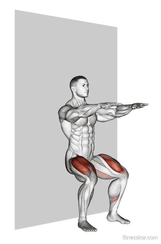
Incorporating heavy isometric knee extensions or isometric wall sits at the very beginning of your leg training acts as an evidence-based "neurological reset." It downregulates tendon pain, clears neuromuscular inhibition, and primes your quadriceps to fire with maximal power during subsequent compound movements.
- Sets
- 4–5
- Reps
- 45s hold
- Rest
- 90 s
Movement Mechanics: Busting the "Knees Past Toes" Myth
One of the most persistent dogmas in commercial fitness is the warning that your knees must never travel past your toes during a squat or lunge. This outdated myth originated from a flawed 1978 study that observed increased patellofemoral shear stress when the knees drifted forward. While the study's mathematical observation of compressive force was correct, its practical conclusion was disastrously wrong.
When you artificially restrict forward knee translation during a squat, you do not eliminate mechanical stress; you simply transfer it up the kinetic chain into your lumbar spine and hip joints. Biomechanical analyses confirm that forcing a vertical shin angle increases hip torque by over one thousand percent, forcing the torso to fold forward and placing immense shear stress across the L4–S1 spinal segments.
More importantly, your knees are anatomically designed to travel past your toes. Every time you walk down a flight of stairs, hike downhill, sit into a chair, or perform an Olympic clean, your knees must translate forward beyond your forefoot. If you never expose your knees to deep flexion under load in the gym, your patellar tendons and cartilage never develop the structural tolerance required for real-world athletic demands.
To squat deeply without knee irritation, the real key is not restricting knee travel, but ensuring that knee flexion occurs in alignment with adequate ankle dorsiflexion and strong hip stabilization. If your ankles are stiff and unable to dorsiflex freely, your body will compensate by collapsing into dynamic knee valgus (inward caving), which concentrates shear forces directly onto the medial patellar facet and MCL. For lifters working through ankle restrictions, our companion guide on hip mobility guide and dedicated ankle mobility drills provide the missing foundation.
The VMO and Scapular-Pelvic Synergies
A durable knee cannot exist in isolation; it functions as the middle link in a closed kinetic chain bookended by the foot below and the pelvis above. The knee is often described as an "innocent bystander" caught between what is happening at the ankle and the hip.
When lifters complain of diffuse anterior knee pain or patellar tracking issues, the underlying dysfunction frequently traces back to an imbalance between two muscle groups:
- The Vastus Medialis Oblique (VMO): The teardrop-shaped inner head of the quadriceps. The oblique orientation of its fibers pulls the patella medially, directly counteracting the lateral pull of the much larger vastus lateralis and the iliotibial (IT) band. When the VMO is weak, inhibited, or delayed in its firing sequence, the patella tracks laterally out of its physiological groove, grinding against the lateral femoral condyle.
- The Gluteus Medius and External Rotators: The lateral hip muscles control the transverse and frontal plane alignment of the femur. When the gluteus medius fails to stabilize the pelvis during single-leg stance or heavy squatting, the femur internally rotates and adducts. This dynamic valgus collapse dramatically increases the "Q-angle" (quadriceps angle), magnifying lateral displacement vectors on the patella and putting tremendous strain on the patellar tendon.
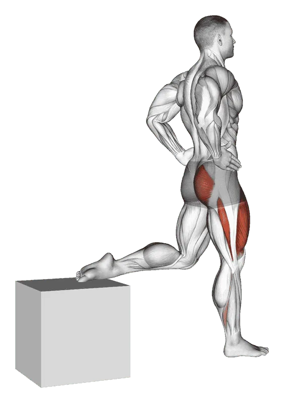
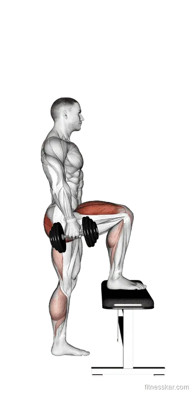
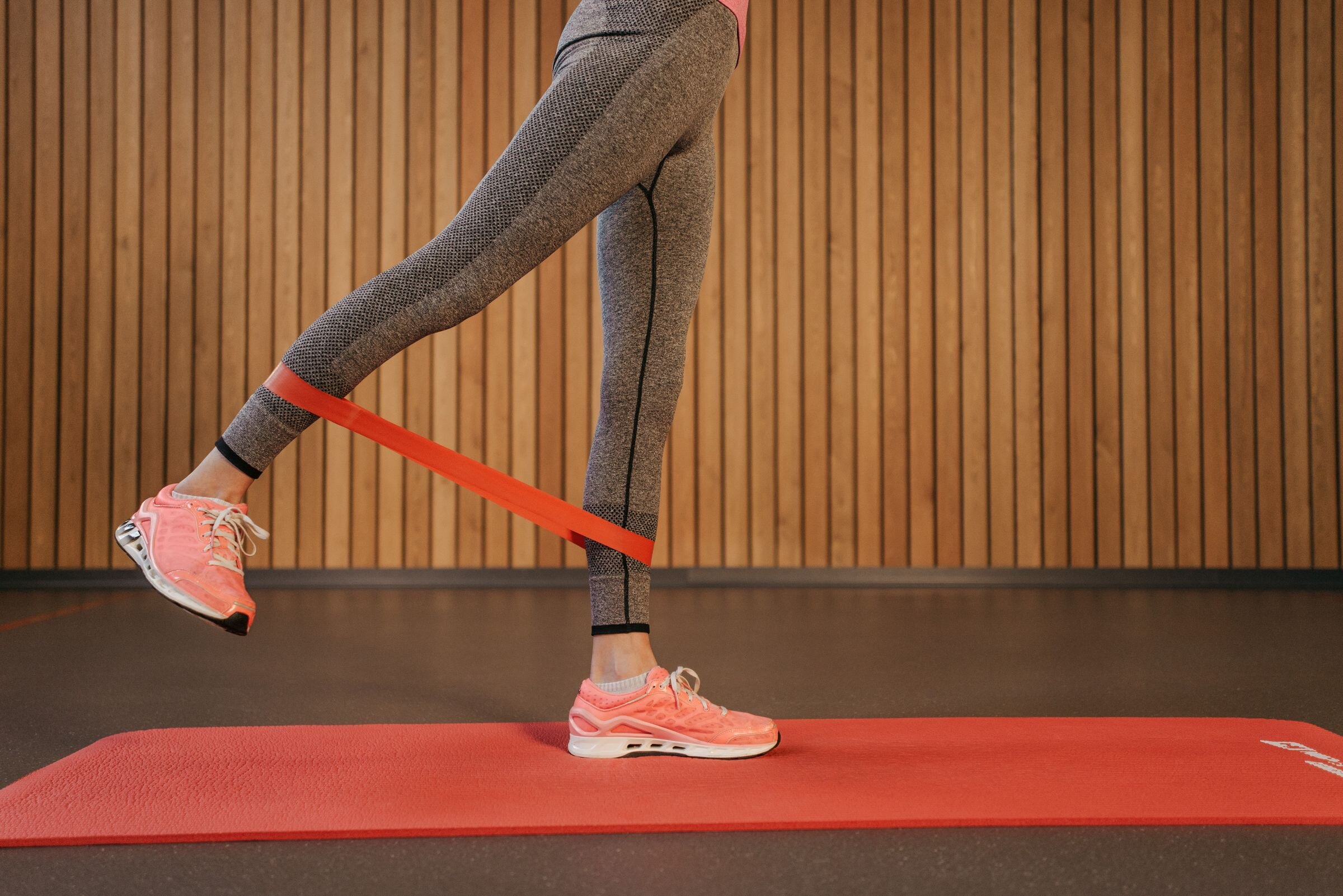
To eliminate patellar tracking pain permanently, you must strengthen terminal knee extension to fire the VMO while simultaneously building robust lateral hip stability. Incorporating movements like Bulgarian split squat and dumbbell step-up forces each leg to stabilize independently, eliminating bilateral compensations and teaching the hip and knee musculature to fire in perfect synchronization.
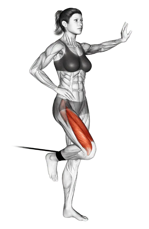

- Sets
- 3–4
- Reps
- 15–20
- Rest
- 60 s
- Tempo
- 2–1–2
The 4-Phase Bulletproof Knee Protocol
Building indestructible knees does not happen by accident; it requires a structured, periodized protocol that progresses from pain modulation to dynamic resilience. Whether you are recovering from mild patellar tendinitis or seeking to safeguard your joints against future injury, follow this four-phase system.
Phase 1: Analgesia and Isometric Loading (Weeks 1–2)
The objective of Phase 1 is to calm down irritated tendons, relieve pain, and eliminate neuromuscular inhibition. Dynamic heavy jumping and deep bouncing squats are temporarily paused in favor of static positions:
- Primary Movement: Heavy Spanish Squats or Wall Sits (wall sit) holding at a seventy- to eighty-degree knee flexion angle.
- Dosing: 4 to 5 sets of 45-second holds, performed with at least 90 seconds of rest between sets.
- Frequency: Can be performed 3 to 4 times per week, or as a pre-workout warm-up prior to lower-body lifting sessions.
Phase 2: Slow Eccentrics and Terminal Extension (Weeks 3–5)
Once resting tendon pain has subsided, Phase 2 introduces controlled mechanical tension to stimulate collagen synthesis and lengthen muscle-tendon units under load:
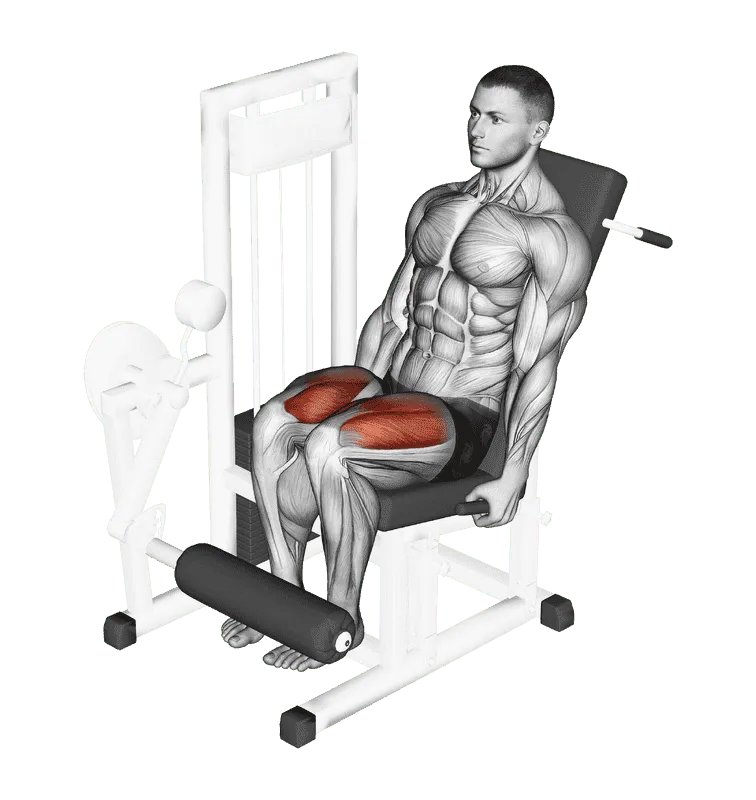
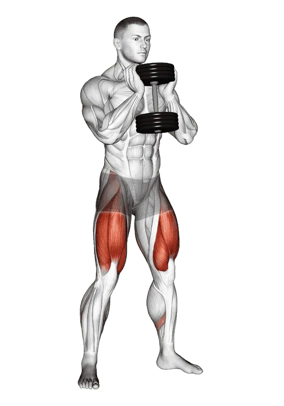
- Primary Movements: Banded Terminal Knee Extensions (band standing leg extension), slow tempo lever leg extension with a 3-second eccentric phase, and slant-board or heel-elevated goblet squats (goblet squat).
- Dosing: 3 to 4 sets of 10 to 12 repetitions utilizing a 3-1-1 tempo (3 seconds lowering, 1-second pause, 1 second lifting).
- Goal: Drive cellular mechanotransduction without inducing sharp tendon flare-ups.
Phase 3: Unilateral Strength and Hip Integration (Weeks 6–8)
Phase 3 bridges the gap between isolated joint health and athletic movement by loading the knee unilaterally across varied planes:
- Primary Movements: Bulgarian split squat, forward and reverse lunges, and controlled dumbbell step-up onto a low-to-medium box with intentional forward knee tracking.
- Dosing: 3 to 4 sets of 8 to 10 repetitions per leg.
- Goal: Correct side-to-side strength deficits, challenge pelvic stability, and build confidence in single-leg deceleration.
Phase 4: Full-Range Dynamic Compound Loading (Week 9 and Beyond)
In the final phase, you integrate your newly acquired joint resilience back into heavy compound barbell lifts, loading through complete anatomical ranges:
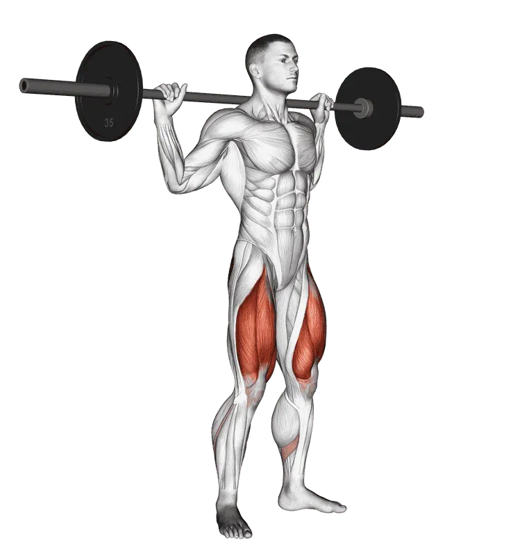
- Primary Movements: Full-depth barbell squat, front squats, and deadlift variations.
- Dosing: Programmed within your primary hypertrophy or strength routine, maintaining deliberate eccentrics and avoiding abrupt bottom bounces.
- Integration: Pair this work with our comprehensive how to squat technique blueprint to cement proper biomechanical cues.
| Phase | Primary Focus | Key Exercise | Sets & Reps | Tempo | Rest Interval |
|---|---|---|---|---|---|
| Phase 1: Analgesic | Pain relief & cortical disinhibition | Wall Sit (exercise:740) | 4–5 × 45s hold | Static hold | 90 seconds |
| Phase 2: Remodeling | Collagen synthesis & VMO activation | Band Leg Extension (exercise:1040) | 3–4 × 15–20 | 2-1-2 | 60 seconds |
| Phase 3: Unilateral | Hip-knee tracking & pelvic stability | Bulgarian Split Squat (exercise:1633) | 3–4 × 8–10 / leg | 3-1-1 | 90 seconds |
| Phase 4: Compound | Full-range structural durability | Barbell Full Squat (exercise:45) | 3–5 × 5–8 | 2-1-1 | 120–180s |
Long-Term Programming for Joint Longevity
Maintaining healthy knees across decades of hard training is not a temporary six-week rehabilitation project; it is an ongoing training philosophy. To keep your joints resilient while pushing for continuous muscle mass and strength gains, integrate the following habits into your weekly programming:
- Manage Rate of Force Development (RFD) Transitions: Tendons are highly sensitive to the speed of movement. While slow, heavy lifting strengthens tendons, rapid ballistic movements (plyometrics, sprint decelerations, jumping) subject tendons to intense elastic shock waves. When introducing explosive drills, progress volume gradually over months rather than weeks.
- Respect the Seventy-Two-Hour Remodeling Cycle: Research into collagen metabolism shows that after heavy tendon loading, collagen degradation spikes before collagen synthesis overtakes it around thirty-six to forty-eight hours post-workout. Net collagen accumulation peaks around seventy-two hours. Training heavy knee-dominant patterns every single day keeps tendons in a perpetual state of net degradation. Allow at least forty-eight to seventy-two hours between intense lower-body sessions.
- Balance the Posterior Chain: For every set of knee-dominant quadriceps work you perform, ensure equal volume for the posterior chain. Strong, active hamstrings and glutes act as an essential braking mechanism, decelerating forward tibial translation and sparing the knee ligaments. Review our how to Romanian deadlift guide to optimize your posterior chain mechanics.
- Utilize Structured Splits: Pairing high-intensity leg sessions with structured recovery days is the cornerstone of sustainable training. Our proven four-day mass routine provides an ideal frequency balance, spacing lower-body training to optimize both muscular hypertrophy and joint recovery. For deeper strategies on tendon health across all major joints, explore our complete joint durability guide.
Warning Signs and When to Seek Medical Guidance
While dull tendon achiness and mild muscular fatigue are common responses to hard training, certain symptoms indicate acute structural pathology that requires formal medical evaluation. Never attempt to "train through" mechanical joint instability.
Frequently Asked Questions
Frequently asked questions
Should I wear knee sleeves for knee pain?
Neoprene knee sleeves provide joint warmth, mild compression, and enhanced proprioceptive feedback, which can make knees feel more comfortable during squats. However, sleeves are passive tools that do not correct tendon pathology or muscular imbalances; use them to support your warm-up, not as a replacement for progressive loading and mobility.
Is walking or cycling good for knee recovery?
Yes. Low-impact cyclic movement promotes synovial fluid circulation throughout the joint capsule without subjecting tendons to high peak forces. Ten to twenty minutes of easy cycling or backward treadmill walking is an outstanding method to warm up the knees and accelerate post-workout recovery.
How long does it take for an irritated patellar tendon to heal?
Mild reactive tendinopathy can calm down within two to four weeks with proper isometric loading and load management. Chronic degenerative tendinopathy requires consistent, progressive tendon remodeling over twelve to twenty-four weeks to synthesize new collagen fibers and restore full load capacity.
Can deep squats damage your knee cartilage?
In healthy, uninjured knees, full-depth squats do not damage cartilage; in fact, the cyclic compressive loading stimulates cartilage hydration, thickens articular surfaces, and strengthens bone mineral density. Cartilage wear occurs primarily when lifters use poor technique, overload beyond their capacity, or force depth through restricted joints.
Sources
- Rio E, Kidgell D, Purdam C, et al. Isometric exercise induces analgesia and reduces inhibition in patellar tendinopathy. British Journal of Sports Medicine. 2015. doi.org/10.1136/bjsports-2014-094386 ↩